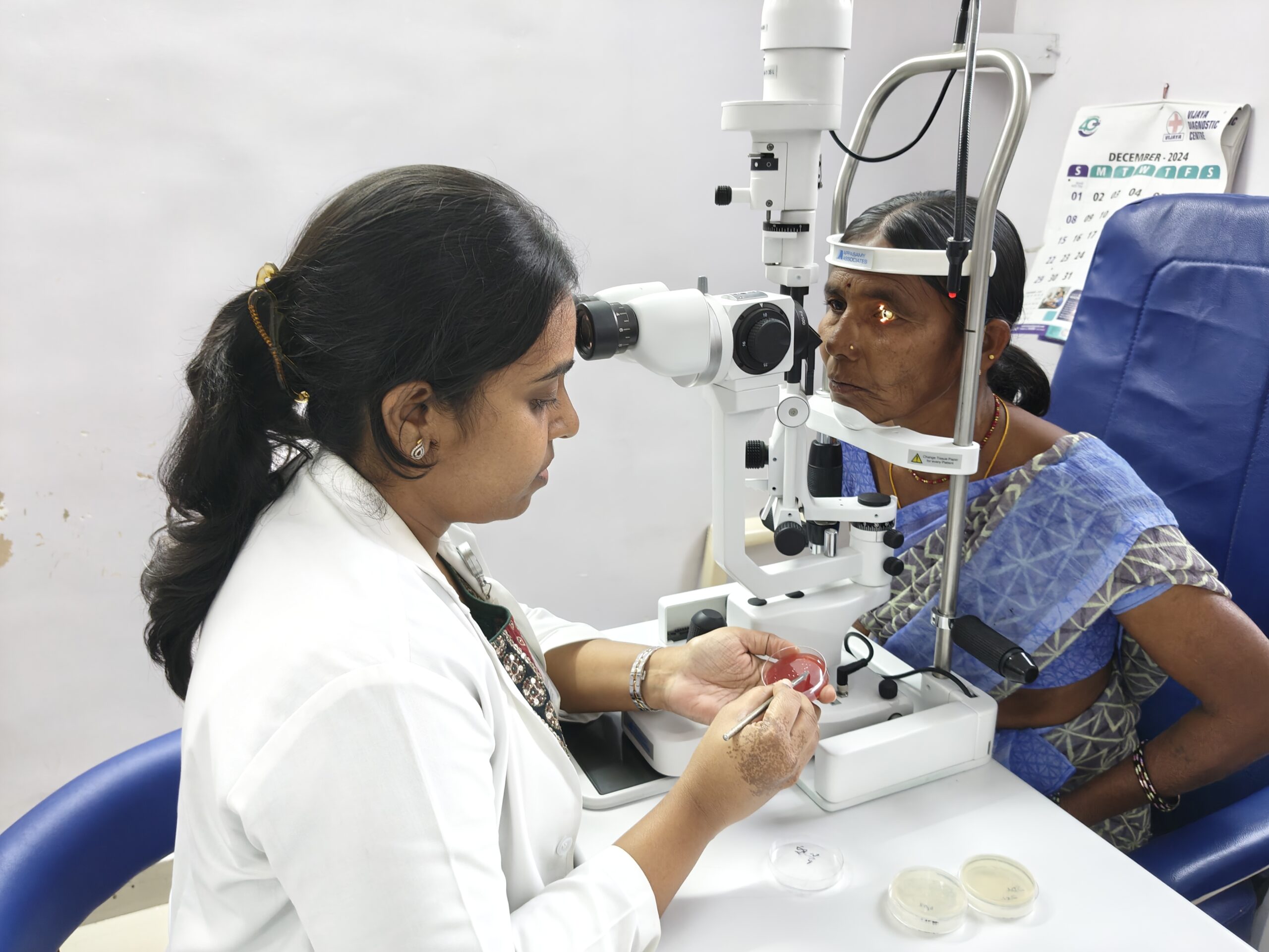
Managing microbial keratitis in resource-limited settings

Related content
In resource-limited settings, the management of corneal infections (microbial keratitis) is challenging. Patients often delay seeking medical help, arriving in clinic with advanced disease, and diagnostic laboratory support may be unavailable.
PART 1 General management principles
In this section, we discuss the general principles of managing a patient with microbial keratitis.
Patients with microbial keratitis need:
1. Early and appropriate diagnosis and treatment, ideally based on microbiology results
2. Good pain management
3. Good adherence with treatment during the first few days (usually achieved by admitting the patient)
4. Regular clinical review.
Early diagnosis and treatment
If there are clinical signs or symptoms to suggest microbial keratitis or corneal infection (see poster on pages 13-14), start patients on a broad-spectrum topical antibiotic and refer them urgently to a specialist eye clinic.
Once reviewed in hospital, most patients can be effectively managed with intensive topical treatment alone, but there are some patients for whom supportive or adjunctive treatment may be needed.
Ideally, the treatment given should be based on microbiological identification of the microorganism involved. In the absence of microbiological services, we suggest you refer to the article on page 3 to guide your decision making. In settings where fungal keratitis is known to be more common than bacterial keratitis, first-line treatment should include antifungal eye drops. If there is any uncertainty about the pathogen responsible, it is advisable to treat empirically with both antifungal and antibiotic eye drops, until a definitive diagnosis can be made.
For most patients with microbial keratitis, treatment typically follows an intensive ‘sterilisation’ phase for up to a week, where drops are instilled every hour. This is followed by a ‘healing’ stage, where the intensity of the drops is reduced to allow the corneal epithelium to heal. The timings and antimicrobial eye drops vary depending on the causative microorganisms.
For more information on the different medical treatment needed for bacterial, fungal and Acanthamoeba keratitis, see Part 2.
Pain management
Microbial keratitis causes severe pain. It is therefore important to manage patients’ pain, as pain is often one of the reasons a patient may start using traditional eye medications or inappropriate conventional medicines, such as topical steroids. We recommend cycloplegia (with topical cyclopentolate or atropine), along with oral painkillers such as paracetamol, codeine, or NSAIDs such as ibuprofen.
Adherence and counselling
Management of microbial keratitis requires a multi-disciplinary approach. It is vital to counsel the patient about the disease, its management, the expected outcome, and the need for long-term follow-up.
Many patients do not get adequate information about how to continue their treatment after being discharged, such as how often to instil their eye drops and how to position themselves to ensure the drops go into the eye. This contributes to inadequate adherence with medication.
Long-term follow-up (of about 2–3 months) is often needed; if patients do not realise the importance of this from the outset, they frequently miss future appointments, which can lead to adverse clinical outcomes.
Monitoring and follow-up
Follow-up is essential to evaluate the effectiveness of prescribed medication in patients with microbial keratitis.
Follow-up frequency depends on disease severity; for most patients a review at 48–72 hours is recommended when initial symptomatic improvement (typically a reduction in pain and discharge) can be observed, along with reviewing any initial microbiology results. Patients with severe microbial keratitis may require admission (if available) and daily monitoring until stable, due to the risk of corneal perforation. If patients have to travel from far away, they should be admitted for as long as possible, ideally for the first few days of treatment at a minimum.
Signs that the patient is getting better include reduced pain, reduced redness and anterior chamber activity, and a reduction in the size of the epithelial defect. However, in fungal keratitis (and in certain forms of bacterial keratitis, such as Pseudomonas keratitis), there can be an initial worsening of clinical signs due to the inflammatory response to dead or dying pathogens. Drug toxicity may also delay the healing process, which requires close monitoring. A reduction in pain and eyelid swelling may be the only initial positive findings.
If patients are not improving, then there are several questions to consider. First, what is their treatment adherence like? Are they using the drops as prescribed? Second, do they have an underlying (often unknown) systemic condition that is responsible, such as uncontrolled diabetes or HIV? Third, is this a mixed bacterial and fungal infection? Finally, are they using traditional eye medicine that is making things worse?
PART 2 Medical treatment
Treatment can be broadly categorised into medical and surgical options. Medical management typically involves the use of topical antimicrobial medication. When medical management is insufficient, or when there are complications, surgical interventions such as corneal debridement, therapeutic keratoplasty, or conjunctival flaps may be needed to preserve vision and the structural integrity of the eye.
In this section, we discuss the medical management of bacterial, fungal, and Acanthamoeba microbial keratitis in a low-resource setting.
A note about corticosteroids
The adjunctive use of topical corticosteroid therapy in microbial keratitis remains hotly debated. While corticosteroids may suppress inflammation and reduce scarring and associated visual loss, potential disadvantages include infection recurrence, local immunosuppression, corneal melting risk, and increased intraocular pressure. Patients already on corticosteroids should reduce or stop them until the infection is under control.
Topical corticosteroids may be considered for patients with severe bacterial keratitis with signs of significant acute inflammation, and if there is clinical improvement following at least 48 hours of intensive topical antibiotics. The dosing frequency should be the minimum amount to control the inflammation.
Steroids should not be prescribed for fungal, Acanthamoeba or Nocardia corneal infections.
Other adjunctive therapy
Oral antibiotics are not usually required unless there is a corneal perforation, when an oral fluroquinolone can be used, e.g. moxifloxacin. There may be a role for oral doxycycline 100 mg once daily to help limit corneal melting and scar formation, particularly in patients with more severe disease.
For patients with deep fungal keratitis, or where there is failure to respond to initial natamycin 5% treatment, oral antifungals such as voriconazole or itraconazole may be given. However, these drugs have dangerous side effects including liver toxicity and therefore should only be given where liver function testing can be performed. Additional treatment for these more challenging, deeper fungal infections includes injections of amphotericin B or voriconazole into the anterior chamber (intracameral injections), or intrastromal around the infiltrate. However, there is little convincing evidence to support the use of intrastromal injections.
Bacterial keratitis
It is always important to base antibiotic choice on local antimicrobial susceptibility profiles if such data are available.
Ideally, admit the patient initially to ensure that the eye drops are administered as planned and to allow regular follow-up. Give treatment as per the frequency in Table 1.

Single-drug therapy with topical fluoroquinolones is as effective as combination therapy (see below). Fluoroquinolone options include ciprofloxacin, ofloxacin, levofloxacin, moxifloxacin and gatifloxacin.
Combination therapy – using a combination of topical cephalosporin (e.g., cefuroxime) and topical gentamicin – can be considered in patients with central or severe keratitis (generally defined as an infiltrate of greater than 2 mm or more), particularly in patients with a hypopyon, or in patients who are unresponsive to initial single-drug therapy with fluoroquinolone.
Ointments are less effective, due to poor corneal penetration, but may be used adjunctively or at bedtime in patients with milder infection. Subconjunctival injections may be considered for patients with adherence difficulties or if there are delays in obtaining fortified antibiotic eye drops. Systemic therapy is reserved for patients with scleritis, endophthalmitis, microbial keratitis associated with systemic infections, or gonococcal keratitis.
Note: In patients with bacterial keratitis (particularly if caused by Pseudomonas spp.), the clinical signs can often be worse in the first few days after starting treatment; for example, the size of the hypopyon may increase. However, if the patient’s pain is reducing significantly, this is a reassuring sign that the treatment is working. Treatment should then continue unchanged, and the patient be reviewed further in a few days’ time.
Fungal keratitis
The standard treatment for fungal keratitis is natamycin 5% drops. The treatment schedule is given in Table 1.
Voriconazole 0.1% drops may be considered if natamycin is not available, although it may be prohibitively expensive. In the absence of commercially produced antifungal eye drops, 0.2% chlorhexidine drops can be considered an alternative treatment. Chlorhexidine has the advantage of being inexpensive and easy to formulate (see article in issue #118 www.cehjournal.org/articles/285).
Amphotericin B 0.15% eye drops have also been used in recalcitrant cases of filamentous fungal keratitis, and remains first-line treatment for patients with non-filamentous Candida keratitis. This could be made in a hospital, as per the instructions in the panel.
Amphotericin B 0.15% eye drop preparation
What you need:
• A 50 mg vial of liposomal amphotericin B parenteral powder for injection (the type used to constitute an IV drip; brands from India include Amfocare)
• Distilled, sterilised water
• Artificial tears (eye drops)
• Sterile eye drop bottle (minimum 10 ml).
Method
• Mix the 50 mg vial of liposomal amphotericin B parenteral powder with 10 ml of distilled, sterilised water. It should fully dissolve without any precipitates.
• Add 3 ml of this preparation to the eye drop bottle, followed by 7 ml of artificial tears (eye drops).
• Store the solution at 4 degrees Celsius; it can be used for 1 week.
While there is an epithelial defect, use a topical broad-spectrum quinolone antibiotic (such as moxifloxacin, ciprofloxacin, or ofloxacin, depending on local availability and resistance patterns) to prevent a secondary bacterial infection. This is administered four-times per day until the epithelium heals.

As with bacterial infections, the patient should be admitted initially to ensure the eye drops are administered as planned and to allow regular follow-up.
Acanthamoeba keratitis
Acanthamoeba keratitis poses a challenge in treatment due to its resistant cyst stage, which is less responsive to therapeutic agents compared to the trophozoite stage. Biguanides, particularly chlorhexidine and polyhexamethylene biguanide (PHMB), are commonly used drugs, either alone or in combination with diamidines.
PHMB 0.06-0.08% monotherapy can be used as first-line treatment for Acanthamoeba keratitis. However, in resource-limited settings where fungal keratitis is also prevalent and PHMB may not be available, it is pragmatic to use chlorhexidine (typically 0.02%, but 0.2% can be used if that is all that is available), ideally in combination with a diamidine such as propamidine isethionate 0.1% and hexamidine 0.1%. However, prolonged use of propamidine isethionate may lead to toxic keratopathy, as well as iris atrophy, cataract, and peripheral ulcerative keratitis.1
Initial therapy for Acanthamoeba keratitis involves frequent administration (Table 1), gradually tapering based on response, with treatment durations ranging from 3 months to over a year.

Management of complications secondary to microbial keratitis
Despite appropriate treatment, some patients with microbial keratitis develop complications including descemetoceles (exposure or protrusion of Descemet’s membrane) and corneal perforations. For some patients, the cornea continues to melt. The recommended treatment for some of these complications would be to do an amniotic membrane transplantation, seal the perforation using tissue glue, use a bandage contact lens, or perform a therapeutic keratoplasty. These are, however, not available in many low-resource settings. In resource-limited facilities, small perforations and descemetoceles can be managed using procedures like conjunctival flaps (Figure 1) and temporary tarsorrhaphy, or surgical eyelid closure. The use of conjunctival flaps in patients with active corneal infection is not encouraged as it may aggravate the infection. We recommend that these patients first undergo a temporary tarsorrhaphy and, once there are no signs of infection (negative microscopy), the conjunctival flap is done. The drawstring procedure of temporary tarsorrhaphy (Figure 2) is recommended, because it enables us to loosen the sutures to examine the eye without the need for repeat surgery. See issue#89 for detailed guidance on how to perform drawstring tarsorrhaphy: www.cehjournal.org.articles/584.

Perforations can be sealed using autologous Tenon grafts for perforations of up to 5 mm or scleral grafts (preferably) for peripheral corneal perforations. Tenon grafts use tissue from the patient’s own eye to treat corneal perforations. The tissue is harvested from the Tenon’s capsule: a dense, elastic, fibrous connective tissue in the eye. Scleral grafts use the scleral rim from a corneal donor and can be stored on the shelf in 100% ethanol. They offer structural support and there is little chance of rejection.

References
1. Dart JKG, Papa V, Rama P, Knutsson KA, Ahmad S, Hau S, Sanchez S, et al. The Orphan Drug for Acanthamoeba Keratitis (ODAK) Trial: PHMB 0.08% (Polihexanide) and Placebo versus PHMB 0.02% and Propamidine 0.1. Ophthalmology. 2024;131(3):277-287. DOI: 10.1016/j.ophtha.2023.09.031.
